Sunday, March 23, 2014

sympathetic system: adrenergic receptors in human body

receptor for sympathetic system we called adrenergic receptors which are divided into 2 families: a receptors and b receptors.

a receptors:

  1. a1 receptors:
  • vasoconstriction : increase peripheral resistance and blood pressure
  • glycogenolysis, gluconeogenesis in liver
  • increase tone of sphincters in git and bladder
  • decrease tone oof motility of git
  • contraction of pregnanat uterus
  • mydriasis by contraction of dilator pupilla muscle.
    2. a2 receptors:
  • presynaptical localization: result in decrease release of noradrenaline
  • postsynaptical localization: decrease insulin release by b cell, decrease motility , tone and secretion in git
  • aggregation of platelets
  • vasoconstriction
  • inhibit acetylcholine release
b receptors:

1. b1 receptors:
  • positive effect on heart: increase heart rate, contractility, dromotropic effect(conductivity), bathmotropic(excitibility)
  • kidney-juxtaglomerular : increase renin secretion
2. b2 receptors :
  • vasodilation of blood vessels in skeletal muscles and coronary arteries.
  • bronchodilation
  • relaxation of visceral smooth muscle
  • increase muscle and liver glycogenolysis
  • increase insulin release by b cells
  • increase glucagon release
  • increase salivation-thick
  • decrease tone of bladder detrussor muscle
  • decrease tone of uterine smooth muscle
  • skeletal muscle tremor
3. b3 receptors
  • lipolysis

Saturday, March 15, 2014

grow new teeth using ultrasound transducer

20 minutes a day for four months to stimulate growth of a tooth’s root
Nanotechnology expert Dr. Jie Chen and his associate Dr. Ying Tsui from the Faculty of Engineering at the University of Alberta have developed a new ultrasound transducer to stimulate the growth of teeth and fix asymmetric jaw bones. The transducer utilizes LIPUS, which stands for “low-intensity pulsed ultrasound.” The miniature, wireless device does exactly what its name suggests: it sends pulses of ultrasound into biological matter, such as gums, muscles or bones to increase healing or stimulate growth of new tissue.
The transducer is a type of “system-on-a-chip,” which generally refers to an incredibly small system, like a microchip, that contains all the parts and electronic circuitry a system needs to run. Advances in system-on-a-chip technology allow scientists and researchers the opportunity to work on an increasingly smaller scale with computing power akin to computers from around a decade ago.
Perhaps the best part, other than growing new teeth, is the transducer doesn’t need to be surgically implanted. A patient need only apply the device topically for 20 minutes a day for four months to stimulate growth of a tooth’s root. So far it has been tested on 12 Canadian patients in the prototype stage. The team hopes to release a commercial form of the technology within the next two years.

Friday, March 14, 2014

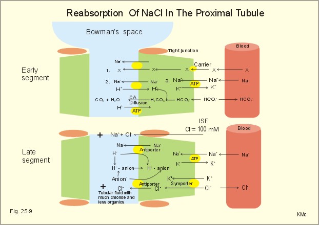
Tubular function in the kidneys


Proximal convulated tubule(PCT)

-          Approximately 120ml/min of glomerular filtrate are generated in glomerulus.
-          In the PCTmost  of the filtered load reabsorb again which are water by osmotic activity (80%) most of the Na and under normal condition almost all K+ by passive diffusion(driven by  the positive tubule electrical potential present along the S2 and S3 segments and by paracellular solvent drag and glucose actively co transpot with Na from the tubular fluid.  Also have Na-H antiporter.
-          Number of substances also secreted by into tubular fluid for example creatinine, histamine many drugs and toxic.

Loop of Henle
-          Normally about 30ml/min of isotonic filtrate is delivered to loop oh Henle where counter multiplier mechanism can achieves concentration of the urine.
-          Loop of Henle important to make sure osmotic gradient for facultative water reabsorption from collecting tubule to prevent water loss by kidney. Along the descending limb of the loop of Henle, K is secreted into the tubule lumen from the interstitium. Along the thick ascending limb, K is reabsorbed via Na-K-2 Cl cotransport. In the loop, there is net K reabsorption of 25% of the filtered K. Na reabsorption is controlled by sympathetic system and aldosterone.

Distal convulated tubule(DCT)

-          In the distal convulated tubules there are facultative N and Ca reabsorption and K and P excretion controlled by aldosterone and parathyroid hormone.

Collecting duct
-          It contain 2 types of cells: principal cell and intercalated cell.
-          The principal cell mediates the collecting duct's influence on sodium and potassium balance via sodium channels and potassium channels located on the cell's apical membrane. Aldosterone determines expression of sodium channels with increased aldosterone causing increased expression of luminal sodium channels. Aldosterone increases the number of Na⁺/K⁺-ATPase pumps  that allow increased sodium reabsorption and potassium secretion. Vasopressin determines the expression of aquaporin channels on the cell surface. Together, aldosterone and vasopressin let the principal cell control the quantity of water that is reabsorbed.
-          Intercalated cells come in α and β varieties and participate in acid-base homeostasis
Type of cell
Secretes
Reabsorbs
α-intercalated cells
acid (via an apical H+-ATPase and H+/K+ exchanger) in the form ofhydrogen ions
bicarbonate (via band 3, a basolat-eral Cl-/HCO3-exchanger)[5]
β-intercalated cells
bicarbonate (via pendrin a specialised api-cal Cl-/HCO3-)
acid (via a basal H+-ATPase)
For their contribution to acid-base homeostasis, the intercalated cells play important roles in the kid-ney's response to acidosis and alkalosis.

Thursday, March 13, 2014

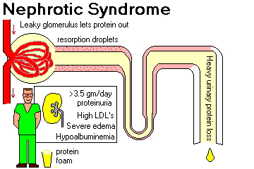
nephrotic syndrome

What is nephrotic syndrome?

Nephrotic syndrome caused by increase in glomerular permeability to plasma protein. This syndrome can arise from nephritc syndrome when severe proteinuria occur which is daily loss of protein through kidneys is more than 3.5g/day.
Usually loss of proteins more than production by liver causing hypoproteinemia. When there is hypoproteinemia, oncotic pressure in the blood reduced causing formation of edema , edema usually can be detected in infraorbital region and lower limbs. Because of the body has lower protein concentration, liver will try to produce more protein but, there is not enough protein in the body causing liver produce lipoprotein resulting in hyperlipidemia usually formation of LDL, and cholesterol more than 10mmol/L. hyperlipidemia can cause increase risk of atherosclerosis.

Besides that , when there is low protein in the body , the substrate to produce immunoglobulin will decrease causing decrease resistance to infections.

Other than that, hypoproteinemia causing loss of anti coagulation factors resulting in hypercoagulability.

Monday, March 10, 2014

cystic fibrosis

Cystic fibrosis also known as mucoviscidosis because this disease cause formation of viscous mucus in lungs, pancreas and also testes.

This disease is hereditary autosomal recessive disease. cystic fibrosis can cause restriction to breathing because the mucus obstruct the lung and also fill the airspace with mucus that is very hard to remove by coughing.

Cystic fibrosis occur when genetic that codes for CFTR(cystic fibrosis transmembrane regulator) is defect causing formation of abnormal CFTR. Abnormal CFTR cannot regulate the chloride ions out the cells causing chloride ions trap inside the cells. this causing disbalance of electric charge inside and outside the cells causing movement of cation mainly sodium ions into the cells and result in formation of salt. when sodium channel is move into the cells , it cause osmotic pressure changes resulting diffusion of water into the cells resulting in dehydration and thick mucus.

when there is thick mucus in lungs, lungs cannot be cleaned properly causing recurrent infections result in chronic pneumonia and formation of lung fibrosis and finally result in cor pulmonale whic is right heart failure.

Sunday, March 9, 2014

anaphylactic shock

Anaphylactic shock: life threatening allergic reaction

Anaphylactic shock is a type 1 hypersensitive reaction (IgE mediated) which occur systematically which life threatening while asthma also in the same group but only occur locally. When you exposed to allergen for the first time , IgE will produced and bind to mast cells. When you reexposed to the same allergen, especially via injury or food, this cause release of histamine from mast cells to all body regions causing capillary leaks, bronchopspasm, wheeze, cyanosis, edema, hypotension because of loss of fluid by formation of edema, tachycardia and urticaria.

Example of precipitants are drugs for example penicillin, latex, stings, eggs, peanuts and pollens.

How we manage anaphylaxis:

First of all, we need to secure the airways and give 100% O2 intubation if respiratory obstruction. The next step is we need to remove the cause and raising the feet to help restore blood circulation. Then we give adrenaline to restore blood pressure and for brochodilation. For further detail you can refer Wikipedia.

Problem during diving in the sea: nitrogen narcosis & Caisson disease

When you diving into deep sea, this 2 problems will come out, but there are solutions for you.

  1. Nitrogen narcosis occur when you diving more tha 80m, nitrogen gas will dissolve into body tissue especially in fat and also nervous tissue. This cause interference in transfer of signal across the neural synapses.this effect mental, physical and finally cause unconsciousness. But if you want to diving more deeper, you can replace nitrogen with helium, but its still has limitation until 150m. if you go deeper, another complication will occur which is neuromuscular disorder called high-pressure nervous syndrome (HPVS): tremors, vertigo and nausea.
  2. Caisson disease is a decompression disease which occur when you rapid come out from deep water. When you diving for every 10 m, air pressure will increase 100kPa causing increase nitrogen gas dissolves especially in fat. Nitrogen gas metabolize slower than other gases that why when you rapid coming out from deep water, nitrogen bubbles will formed

Saturday, March 8, 2014

future analgesic for migrain and pain killer: Electronic aspirin

Electronic Aspirin

For people who suffer from migraines, cluster headaches, and other causes of chronic, excruciating head or facial pain, the "take two aspirins and call me in the morning" method is useless. Doctors have long associated the most severe, chronic forms of headache with the sphenopalatine ganglion (SPG), a facial nerve bundle, but haven't yet found a treatment that works on the SPG long-term. A technology under clinical investigation at Autonomic Technologies, Inc., (Redwood City, CA) is a patient-powered tool for blocking SPG signals at the first sign of a headache.
Aspirin often used as an analgesic to relieve minor aches and pains, as an antipyretic to reduce fever, and as an anti-inflammatory medication. This system not use aspirin as treatment but electric signal to block the pain.


The system involves the permanent implant of a small nerve stimulating device in the upper gum on the side of the head normally affected by headache. The lead tip of the implant connects with the SPG bundle, and when a patient senses the onset of a headache, he or she places a handheld remote controller on the cheek nearest the implant. The resulting signals stimulate the SPG nerves and block the pain-causing neurotransmitters. 

This is advantage to patients, because if you use real aspirin, it has side effect which are cause gastrointestinal ulcers, increase risk of intracerebral hemorrhage, and other complication such as nausea and so on.

get a balanced diet

What is a Balanced Diet?

Eating a balanced diet means choosing a wide variety of foods and drinks from all the food groups. This is because , different groups of foods have dfiferent nutrients inside them.The goal is to take in nutrients you need for health at the recommended levels because our body need different amount of different nutrients.

our body need carbohydrate for energy, protein for synthesis of connective tissue, energy, amono acids, enzyme. fat for energy and adipose tissue also hormone. we also need to take different fatty acid which is omega 3 and omega 6.


The World Health Organization (WHO) makes the following 5 recommendations with respect to both populations and individuals:

1.       Eat roughly the same amount of calories that your body is using. A healthy weight is a balance between energy consumed and energy that is 'burnt off'.
2.       Increase consumption of plant foods, particularly fruits, vegetables, legumes, whole grains and nuts.
3.       Limit intake of fats, and prefer less unhealthy unsaturated fats to saturated fats and trans fats.
4.       Limit the intake of sugar. A 2003 report recommends less than 10% simple sugars.
5.       Limit salt / sodium consumption from all sources and ensure that salt is iodized.
6.       Other recommendations include:
·         Essential micronutrients such as vitamins and certain minerals.
·         Avoiding directly poisonous (e.g. heavy metals) and carcinogenic (e.g. benzene) substances.
·         Avoiding foods contaminated by human pathogens (e.g. E. coli, tapeworm eggs).


fainting: first aid-CPR

If a person is unresponsive and not breathing you need to start cardio pulmonary resuscitation (CPR) right away. Gasping is not breathing!  By unresponsive I mean appears lifeless, doesn't move or respond when shoulder is tapped. Yell for someone to call Emergency Medical Services (EMS) and get an an automated external defibrillator (AED).  An AED applied fast may restart the heart. Don't worry, AEDs are user friendly.

Understand that when a victim's heart has stopped pumping blood (cardiac arrest), permanent brain damage can begin in four to six minutes, so you must act fast.

Cardiac arrest can strike at any age and may be caused by many conditions, including heart attack, suffocation, allergic reaction, drowning, choking, or electric shock.

Here's How:

  1. Attempt to wake victim. If the victim is not breathing (or is just gasping for breath), call 911 immediately and go to step 2. If someone else is there to help, one of you call 911 while the other moves on to step 2.
  2. Begin chest compressions. If the victim is not breathing, place the heel of your hand in the middle of his chest. Put your other hand on top of the first with your fingers interlaced. Compress the chest at least 2 inches (4-5 cm). Allow the chest to completely recoil before the next compression. Compress the chest at a rate of at least 100 pushes per minute. Perform 30 compressions at this rate (should take you about 18 seconds).If you are not trained in CPR, continue to do chest compressions until help arrives or the victim wakes up.It's normal to feel pops and snaps when you first begin chest compressions - DON'T STOP! You're not going to make the victim worse.
  3.  Begin rescue breathing. If you have been trained in CPR, after 30 compressions, open the victim's airway using the head-tilt, chin-lift method. Pinch the victim's nose and make a seal over the victim's mouth with yours. Use a CPR mask if available. Give the victim a breath big enough to make the chest rise. Let the chest fall, then repeat the rescue breath once more. If the chest doesn't rise on the first breath, reposition the head and try again. Whether it works on the second try or not, go to step 4.If you don't feel comfortable with this step, just continue to do chest compressions at a rate of at least 100/minute.
  4.  Repeat chest compressions. Do 30 more chest compressions just like you did the first time.
  5. Repeat rescue breaths. Give 2 more breaths just like you did in step 3 (unless you're skipping the rescue breaths).
  6. Keep going. Repeat steps 4 and 5 for about two minutes (about 5 cycles of 30 compressions and 2 rescue breaths).If you have access to an automated external defibrillator (AED), continue to do CPR until you can attach it to the victim and turn it on. If you saw the victim collapse, put the AED on right away. If not, attach it after approximately one minute of CPR (chest compressions and rescue breaths).
  7. After 2 minutes of chest compressions and rescue breaths, stop compressions and recheck victim for breathing. If the victim is still not breathing, continue CPR starting with chest compressions.
  8. Repeat the process, checking for breathing every 2 minutes (5 cycles or so), until help arrives. If the victim wakes up, you can stop CPR.


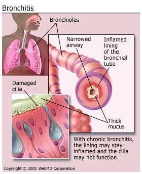
what is chronic bronchitis?

Chronic bronchitis:

defined clinically as cough and sputum production on ost days for 3 months of 2 successive years.symptoms improve if they stop smoking.

How chronic bronchitis develops:

When there is smoking or air pollution, this cause reduce ciliary and pahgocytary activity of epithelium resulting in mucus accumulation.

This lead to bronchi wall inflammation. Inflammatory infiltration of wall lead to edema resulting in obstruction of the airways. Ifnflammation produce exudate result in cough and expectoration. In small bronchioles , this lead to wall destruction eventually bronchiectasis.

When there is obstruction , its result in in prolonged expiration eventually air trapping causing hypoventilation. Hypoventilation result in hypoxia and hypercapnia. Hypoxia and hypercapnia cause precapillary vasoconstriction lead to pulmonary hypertension finally result in cor pulmonale.

Hypoxia also lead to increase production of erythropoietin resulting in polycytemia.
We can detected hypoxia by formation of cyanosis.


Chronic bronchitis also lead to emphysema by chronic inflammation which cause production of proteolytic enzyme that damage the alveoli.

treatment for chronic brochitis is quite the same with asthmatic bronchiole


what is asthma?

Asthma bronchiole

Asthma affect 5-8% of the population. It is characterized by recurrent episodes of dyspnoea, cough usually at night, and wheeze caused by reversible airways obstruction.

The difference between asthma and COPD is that asthma is reversible bronchoconstriction.
When examine patient with astma , pulsus parodoxus may be noted.

Treatment:
1.       Stimulate sympathetic system to open the airways using B agonist.(B2 receptor)
2.       Inhibit parasympathetic systemusing anticholinergic
3.       Reduce inflammation using glucocorticoids.

There are 2 types of astma:
1.       Allergic astma-predominatly occur to children. This results from sensitization to allergens. when there is exposure to allergen,  stimulation of IgE OCCURS. THEN IgE bind to mast cells. When there is next exposure to allergen, it will be detected by mast cells that bind to ige, reeleasing histamine. Histamine cause mucus secretion , inflammation, vascular permeability and bronchospasm and lead to narrowing of airways.

2.       Idiopathic asthma. Usually occur after 40 years old. Usually occur after infections.

Thursday, March 6, 2014

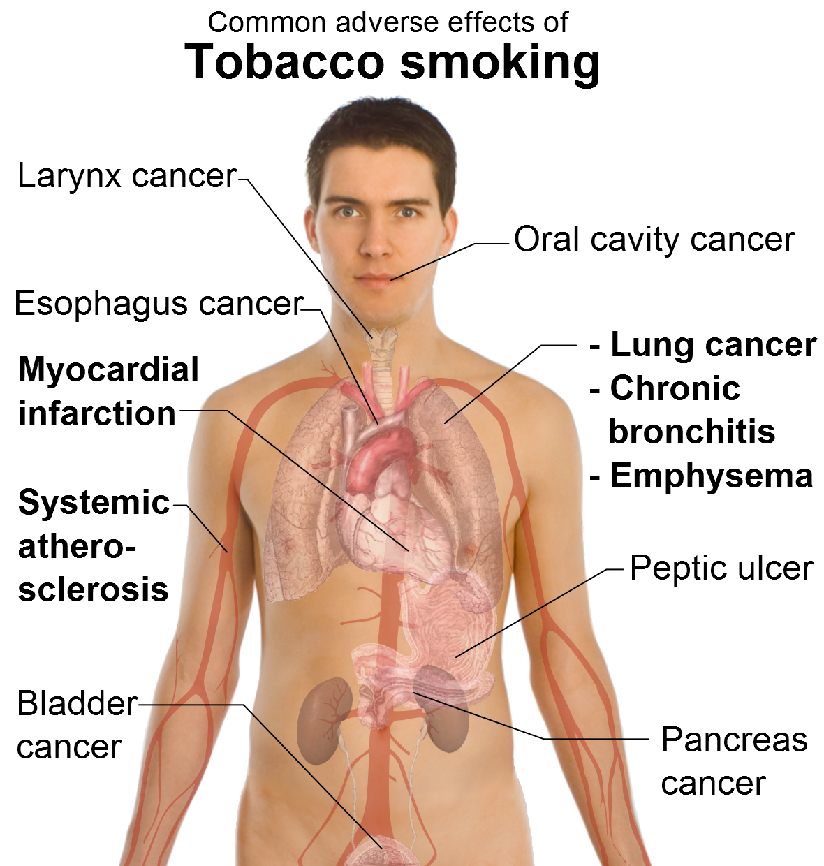
HOW TO STAY HEALTHY? Quit smoking

Dangerous chemicals in tobacco smoke

The most damaging components of tobacco smoke are:

Tar –  The particles contain chemicals, including several cancer-causing substances (carcinogens). Tar is sticky and brown, and stains teeth, fingernails and lung tissue. Tar contains the carcinogen benzo(a)pyrene

Carbon monoxide – this odourless gas is fatal in large doses because it takes the place of oxygen in the blood. Each red blood cell contains a protein called haemoglobin that transports oxygen molecules around the body. However, carbon monoxide binds to haemoglobin better than oxygen because it has higher affinity compare to oxygen. This means that less oxygen reaches the brain, heart, muscles and other organs

Free radicals – these highly reactive chemicals can damage the heart muscles and blood vessels. They react with cholesterol, leading to the build-up of fatty material on artery walls. Their actions lead to formation of atherosclerosis that can effect heray, brain kidney and lower legs.

Metals – tobacco smoke contains dangerous metals including arsenic, cadmium and lead. Several of these metals are carcinogenic and toxic to internal organ for example ti liver and pancreas.

 
HOW TO QUIT SMOKING?

Quit smoking is really difficult because nicotine in cigarette is very addictive. The most important thing is you need to have strong motivation to stop smoking because smoking give you a lot of health problems and reduce quality of life.
You also need to get support especially family and you friends and avoid to be close with friends that smoking.
Besides that , always do exercise and drink a lot of water.
In certain pharmacy, you can find nicotine pills , you can take it and slowly reduce the dose, and finally you can stop nicotine addiction.


HYPOGLYCEMIA

Hypoglycemic states:
Hypoglycaemia is condition we have low glucose concentration than normal in blood. Noemal glucose around 5mmol/L. this cause by  regulation disorder by decrease income of glucose and gluconeogenesis , increase glucose disposal through exercise and hyperinsulinism
It can be :
  •   Fasting hypoglycaemia
  •    Insulinoma
  •  Postprandial hypoglycaemia especially after stomach resections.

What is etiology for hypoglycaemia:
  1. Nutrition disorders

·         This is because of severe fasting and mental anorexia. This cause decrease in substrate offer causing glycogen production and decrease gluconeogenesis.
2                 2.     Conditions based on decrease glucose production

·         Shortage of contra regulatory hormones which are hypothyroidism, renal cortex insufficiency, glucagon defcit and hypothyreosis.
·         Serious liver disease cause decrease amount of glycogen and gluconeogenesis
·         Alcohol excess in undernourished people or after exercise causing inhibition of gluconeogenesis because substrate for glucose formation is use to metabolise alcohol.
·         Enzymopathies.Lack of certain enzyme to synthesis glucose

                  3.  Increase glucose disposal by hyperinsulinemia.

·         This can be congenital insulinemia, infant diabetic mothers(due to insulin hypersecretion as a response to hyperglycemia.
·         Can also cause by dumping syndrome, where food we eat by pass from stomach to small intestine rapidly causing our intestine cannot digest what we eat.
·         Overdose of insulin in diabetic patients.
·         Therapy with sulphonylurea derivates. Cause increase insulin secretion.
·         Insulinoma, which is tumour of B cells.


Clinical picture:
Hypoglycaemia occur when glucose level below 3.3mmol/L, serious below 2.5mmol/L
1  Adrenergic reaction activation:
·         Excessive sweating
·         Trembling
·         Tachycardia
2 Brain hypoglycaemia
·         Fatigue
·         Mental confusion, cognitive disorders
·         Irritability up to seizure, aggresivity, slurred speech
·         Unconsciousness

Therapy:

60ml og 40% glucose oral or intavenous

UNDERSTANDING DIABETES MELLITUS COMPLICATION


here are complications of diabetes mellitus:

Diabetic retinopathy

Damage of of pericyte covering endothelium causing weakening of blood vessels and cause microaneurysm and increase permeability resulting in protein and lipid leakage
Hyperglycemia also increase proliferative processes cause formation of abnormal vessels resulting in haemorrhage and finally cause visual problems up to blindness.

Diabetic nephropathy

Complication of diabetes mellitus :
  1.   Non nephrotic  proteinuria(microalbuminemia)
  2.  Nephrotic syndrome
  3. Renal failure

Proteinuria:
Formation of microalbuminemia results fromm loss of negative charge of basement membrane in glomeruli because AGE(advance glycated endproduct) decrease of heparin sulphate(negative charge)

Nephrotic syndrome:
Which is loss of negative charge and increase of basement membrane pore size due to :
1.       Increase of intraglomerular pressure because of glycosuria, increase glomerular filtration rate and cause hyperfiltration
2.       Imbalance between several growth factors
3.       Non enzymatic glycation of basement membrane constituents

Chronic renal insufficiency:
Diabetes mellitus causing deposition of glucose to extracellular space causing thickening of basement membrane and diffuse proliferation of mesangial cells eventually nodular deposition of hyaline to mesangium.
Gradually mesangial cells infringe on capillary lumen casue decrease glomerular filtration up to complete obliteration result in renal insufficiency.
Diabetic neuropathy

Diabetes mellitus effect nervous system in our bodies.
It cause microangiopathy of small artery that supply nerve fibers. This is because hyperglycemia .
It also cause distal neuropathy where it cause symmetric loss of skin sensitivity resulting in diabetic foot and motor problems.
DM also cause wegetative nerve dysfunction :
1.       Impotention and urine incontenetia
2.       Stomach dystonia and diarhoeas
3.       Anhidrosis(can not sweat properly) or profuse sweating

Macroangiopathy

A disease of the large blood vessels in which fat and blood clots build up and stick to the vessel walls, blocking the flow of blood. Types of macroangiopathy include coronary artery disease (macroangiopathy in the heart), cerebrovascular disease (macroangiopathy in the brain), and peripheral vascular disease (macroangiopathy that affects, for example, vessels in the legs).when formation of artherosclerosis increases, this cause arterial hypertension, coronary heart disease, infarctions, decrease perfusion of the legs result in ischemic changes and neuropathy finally diabetic foot with ulceration and formation of gangrene(black foot)
DM accelerated atherosclerosis by formation of:
  1.   Advanced glycated end products(AGE)
  2.     Dyslipoproteinemia-increase VLDL, cholesterol,LDL, and decrease HDL

AGE:
Formation of AGE is non receptor mediated mechanisms which effect extracellular matrix especially collagen cross linking. Its also enhanced synthesis of extracellular matrix components, trapping of LDJ in the subendothelium and this cause quenches nitric oxide.
Hyperglycemia also modified lipoprotein by glycosylation. Glycosylated LDL has lower recognition by cellular LDL receptor and cause high concentration of LDL in blood and increase susceptibility of LDL to oxidative modification
Its also cause endothelial dysfunction by increasing permeability of endothelial cell monolayers, procoagulant activity, expression of adhesion molecules and intracellular oxidative stress.

Dyslipoproteinemia:
 DM type 1
When there is lack of insulin, reduced inactivation of lipoprotein lipase and increased activity of hormone sensitive lipase causing hyperlipoproyeinemia resulting rapid formation of artherosclerosis.

DM type 2
Concentration of insulin normal at the beginning , when there are certain factor that cause insulin resistance for example inflammation, hyperglycemia or increased free fatty acids in blood, this cause muscle and adipose tissue decrease uptake of glucose resulting in hyperglycemia. When high glucose in blood, this glucose must be converted to glycogen in liver, but when the capacity of liver to store glycogen reached maximum, liver must convert glucose tto lipoproteinemia resulting in hyperlipoproteinemia. Hyperlipoproteinemia increase activity of lipoprotein lipase and decrease activity of hormone sensitive lipase resulting in fat depositon causing obesity. Hyperlipoproteinemia also increase formation of artherosclerosis.